The Library·Reference·Last reviewed September 2026
Eight peptides, read against the evidence
Reference summaries of the preclinical literature: what the animal and cell-culture studies report, at what scale, and where each evidence base thins. Every entry names its species and states its gaps.
Name BPC-157Classification Gastric pentadecapeptidePrimary model species Sprague-Dawley rat
BPC-157
A fifteen–amino-acid fragment of a human gastric protein, studied almost entirely in rat injury models from one Croatian research lineage.
What it is
BPC-157 is a synthetic pentadecapeptide whose sequence derives from a larger protein found in human gastric juice — the “body protection compound” from which it takes its name. It entered the experimental literature in the early 1990s through the work of a Zagreb-based research group interested in cytoprotection, the capacity of certain gastric peptides to limit tissue damage far from the stomach itself. It is not an approved medicine anywhere, and it is not a dietary ingredient; it exists as a laboratory reagent and as the subject of a substantial, unusually single-sourced rodent literature.
What the animal literature reports
The core of the literature is a long series of studies in Sprague-Dawley rats, most of them published across the 1990s, 2000s, and 2010s by the same Zagreb laboratory and its collaborators. In a rat Achilles tendon transection model, investigators reported faster histological and functional recovery in treated animals: earlier collagen organization, greater tensile strength at defined time points, quicker return of gait measures. Related papers described analogous findings in rat medial collateral ligament injury, in muscle crush and laceration models, and across a wide range of gastrointestinal lesion models, from ethanol-induced gastric damage to induced fistulas and anastomotic healing.
The breadth is striking. BPC-157 has been reported in rats to affect angiogenesis, growth factor expression, nitric oxide pathways, and counteraction of various drug-induced toxicities — a range of claims broad enough to warrant caution on its own, since single agents with dozens of distinct reported benefits are rare in pharmacology and should prompt questions about model sensitivity and selective reporting.
Where the evidence thins
Three qualifications dominate. First, single-lab dominance: the overwhelming majority of the rodent work originates from one research lineage. Independent laboratories have published comparatively little, and the finding set has not been stress-tested by groups without a stake in the outcome. Second, methodology: sample sizes are typically small, often single digits per arm, and blinding and randomization procedures are inconsistently described. Third, translation: there are no registered, completed, controlled human trials establishing safety or efficacy for any indication. Findings in rat tendon models are hypothesis-generating. They are not evidence that any effect would occur in human tissue.
Name Thymosin β4 / TB-500Classification Actin-sequestering peptide and synthetic fragmentPrimary model species Mouse, rat
Thymosin β4 and the TB-500 fragment
A ubiquitous intracellular peptide with wound-repair associations in mice, and a synthetic fragment whose name travels further than its evidence.
What it is
Thymosin β4 is a 43–amino-acid peptide found in nearly all mammalian cells, where it binds monomeric actin and helps regulate cytoskeletal dynamics. Its biological role in cell migration made it a plausible candidate for wound-repair research. TB-500 is a synthetic fragment corresponding to a short active region of the parent molecule. The two names are often used interchangeably online; in the published literature they are not interchangeable, because most of the rigorous animal work concerns the full-length thymosin β4, not the fragment.
What the animal literature reports
In murine wound models, full-length thymosin β4 has been reported to accelerate re-epithelialization and increase markers of angiogenesis in treated animals. A widely cited line of mouse cardiac work described improved measures of cardiac function and reduced scar size when thymosin β4 was administered around experimental myocardial infarction, with proposed mechanisms involving epicardial cell activation and neovascularization. Rat corneal injury models have also reported faster closure of epithelial defects.
The actin-sequestering mechanism is well characterized at the biochemical level, which gives the field a coherent molecular story: a peptide that governs cell motility could, in principle, influence how cells migrate into a wound bed. Whether that biochemical story accounts for the tissue-level observations in mice is less settled; wound healing engages dozens of pathways, and the contribution attributable to thymosin β4 specifically is difficult to isolate.
Where the evidence thins
The cardiac findings, though influential, emerged primarily from one laboratory group, and subsequent independent confirmations have been partial. Effect sizes reported in small mouse cohorts tend to shrink when studies are repeated at larger scale. The TB-500 fragment has a far thinner publication record than its parent peptide: much of what is asserted about it traces back to full-length thymosin β4 papers, silently generalized. Human evidence consists of a small number of early-phase studies of the full-length peptide in wound contexts — not enough to characterize benefit, and none of it speaks to the fragment.
FIG. 02 — Back volumes in a research library. Most of what is known about these peptides still lives in print runs few readers ever open.
Name GHK-CuClassification Copper tripeptide complexPrimary model species Rat, mouse; cultured human fibroblasts
GHK-Cu
A plasma-derived copper-binding tripeptide with decades of cell-culture data and a long way still to travel from dish to dermis.
What it is
GHK is the tripeptide glycyl-L-histidyl-L-lysine, first isolated from human plasma in the 1970s by Loren Pickart, who observed that plasma from younger donors better supported the growth of cultured cells. The peptide binds copper(II) ions with high affinity, and most experimental work studies the GHK-Cu complex rather than the peptide alone. It became a staple of wound-healing and skin-biology research, and later a familiar name in cosmetic ingredient lists — a commercial afterlife that has substantially outrun the underlying evidence.
What the animal literature reports
In cultured human dermal fibroblasts, GHK-Cu has been reported to increase synthesis of collagen, elastin, and glycosaminoglycans, and to modulate expression of matrix-remodeling enzymes. Gene-expression studies in cell culture have described broad shifts across hundreds of transcripts, though such breadth makes mechanistic attribution difficult. In rodent wound models — full-thickness excisional wounds in rats and mice — investigators reported faster wound closure and greater collagen deposition in treated animals. Rabbit models appear in some of the older literature.
The fibroblast findings are the most internally consistent part of the literature, in part because cell culture is the easiest system in which to control exposure. Concentrations used in culture, however, bear no simple relationship to what intact skin would ever experience, and this gap is routinely elided in secondary accounts.
Where the evidence thins
The distance between a cultured fibroblast and intact skin is the central problem. Skin is a barrier precisely evolved to keep molecules like copper complexes out; whether topical or injected GHK-Cu reaches dermal fibroblasts at meaningful concentrations in a living animal is a pharmacokinetic question the literature has answered only weakly. Rodent wound studies are small, heterogeneous in endpoint selection, and frequently unblinded. Cosmetic-industry citations of this literature almost never note that gene-expression changes in a dish are not evidence of effect in a face. No adequately powered, controlled human trials establish dermal efficacy.
Name KPVClassification α-MSH C-terminal tripeptidePrimary model species Mouse (murine colitis models)
KPV
The last three residues of α-MSH, carried into cells by a peptide transporter, tested mostly against inflamed mouse colon.
What it is
KPV — lysine, proline, valine — is the C-terminal tripeptide of α-melanocyte-stimulating hormone. Researchers observed decades ago that this fragment retained much of the parent hormone's anti-inflammatory activity in experimental systems while lacking its pigmentary effects, which made it a tidy tool for studying inflammation without the confound of melanocortin signaling on skin. A key mechanistic finding, reported from cell-culture and mouse work in the late 2000s, is that KPV can be transported into intestinal epithelial and immune cells via the oligopeptide transporter PepT1, whose expression increases in inflamed tissue.
What the animal literature reports
The flagship model is DSS-induced colitis in mice: dextran sodium sulfate administered in drinking water damages the colonic epithelium and produces a reproducible inflammatory syndrome with weight loss, bleeding, and histological injury. In this model, investigators reported that KPV-treated mice showed reduced disease activity scores, lower expression of inflammatory cytokines, and less histological damage than controls. Parallel cell-culture work in intestinal epithelial cell lines described reduced activation of NF-κB and related inflammatory signaling. A TNBS-induced colitis model, which produces a different immunological profile, yielded broadly concordant reports.
The PepT1 story gives the literature an unusually clean mechanistic thread: the transporter is upregulated under inflammatory conditions, so the peptide's proposed uptake route is most available exactly where inflammation is present.
Where the evidence thins
DSS colitis is a chemical injury model, not Crohn's disease and not ulcerative colitis; it reproduces inflammation, not the human conditions' chronicity, genetics, or relapsing course. The published mouse studies are small, and much of the work traces to a limited set of laboratories with a focus on melanocortin biology. There are no registered human trials of KPV for inflammatory bowel disease or any other indication. The mechanistic elegance of the PepT1 pathway has been demonstrated in rodents and cell lines; whether the same transport economics hold in human inflamed gut at achievable exposures is unknown.
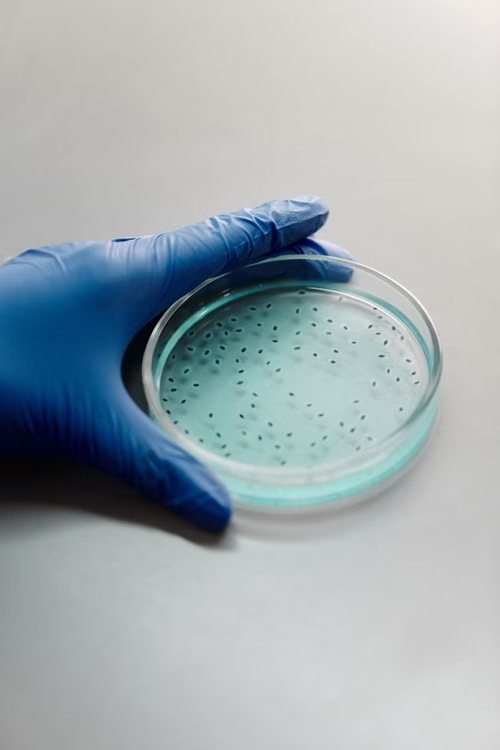
FIG. 03 — A seeded agar plate. Culture systems answer narrow questions well and broad questions badly.
“Replication is not the repetition of a citation. It is the repetition of an experiment, by someone with something to lose.”
The Library, editorial note
Name LL-37Classification Cathelicidin host-defense peptidePrimary model species Mouse; human and murine cell culture
LL-37
The only human cathelicidin: antimicrobial in a dish, immunologically ambidextrous in an animal, and difficult to summarize honestly.
What it is
LL-37 is a 37–amino-acid peptide cleaved from the human cathelicidin precursor hCAP18, produced by neutrophils, epithelial cells, and other immune and barrier tissues. It belongs to the ancient family of host-defense peptides — small, positively charged molecules that disrupt microbial membranes. Unlike most antimicrobial peptides studied as drug candidates, LL-37 is endogenous: human bodies make it constitutively, and its biology is correspondingly tangled, spanning direct microbial killing, biofilm disruption, immune cell recruitment, and modulation of inflammatory signaling.
What the animal literature reports
In mouse infection models, LL-37 and its murine homolog CRAMP have been reported to reduce bacterial burden and improve survival in sepsis and pneumonia paradigms; CRAMP-deficient mice show increased susceptibility to certain skin and intestinal infections, evidence that the peptide family does defensive work in living animals and not only in vitro. Wound models in mice have described impaired healing when the homolog is absent and improved closure when peptide is supplied. Cell-culture studies document activity against a range of bacteria and biofilms at concentrations achievable in laboratory media.
The same literature also reports context-dependent harm: at higher concentrations or in particular tissue environments, LL-37 can promote inflammation, and it has been implicated in the pathology of psoriasis and other inflammatory states in observational human work. Host-defense peptides are regulators, not simple antibiotics. Any honest account of LL-37 has to hold both halves of that sentence at once, which is precisely what makes the peptide so difficult to translate into secondary coverage — and so easy to misquote.
Where the evidence thins
Murine CRAMP is a homolog, not an identical molecule, and mouse immune biology diverges from human in exactly the compartments where LL-37 acts. Antimicrobial potency in broth culture is notoriously a poor predictor of activity in tissue, where salt, serum proteins, and proteases degrade or sequester cationic peptides. Mouse sepsis models have a famously poor record of predicting human sepsis trials. No controlled human efficacy trials establish LL-37 administration as beneficial for any condition, and its double-edged immunology is a reason for caution rather than enthusiasm.
Name Thymosin α-1Classification Thymic immunomodulatory peptidePrimary model species Mouse, rat; immune cell culture
Thymosin α-1
A 28-residue thymic peptide with the deepest literature of any entry here — and a correspondingly complicated relationship with the evidence.
What it is
Thymosin α-1 is a 28–amino-acid peptide originally isolated from thymic tissue in the 1970s during efforts to characterize the thymus gland's influence on immune maturation. It modulates T-cell function and innate immune signaling, including Toll-like receptor pathways. Unlike every other peptide in this Library, a synthetic form is licensed as a medicine in a number of countries for specific viral and oncological indications — a clinical literature that sits outside this publication's preclinical scope and that readers should not conflate with the animal work summarized here.
What the animal literature reports
In mouse models of immunosuppression and infection, thymosin α-1 has been reported to restore aspects of T-cell function, improve pathogen clearance, and improve survival in some bacterial and fungal sepsis paradigms. Rodent tumor models have described enhanced responses when the peptide was combined with chemotherapy or checkpoint-directed approaches, generally framed as restoring immune competence rather than attacking tumors directly. Cell-culture work has mapped effects on dendritic cell maturation and cytokine production.
The mechanistic literature is comparatively mature: receptor-level and signaling effects have been characterized by multiple independent groups, which distinguishes this peptide from most of the Library. That maturity, however, has not resolved the distance between restoring immune markers in a mouse and improving outcomes that matter in a person.
Where the evidence thins
Sepsis is the graveyard of immunomodulatory promise; mouse survival benefits in sepsis paradigms have failed to predict human trial outcomes so reliably that the models themselves are considered part of the problem. Combination-therapy effects in mouse tumor models are sensitive to tumor cell line, timing, and endpoint selection, and many published experiments are small. And while licensed human use exists in some jurisdictions, the strength of the underlying international trial evidence is contested — a reminder that regulatory approval in some countries is not synonymous with demonstrated efficacy by the standards of large, replicated, controlled trials.
FIG. 04 — Histology under review. Endpoint scoring is where unblinded studies most often go wrong.
Name MOTS-cClassification Mitochondrial-derived peptidePrimary model species Mouse (metabolic models)
MOTS-c
A sixteen-residue peptide encoded in mitochondrial DNA, described in 2015, and already carrying more expectation than evidence.
What it is
MOTS-c — mitochondrial open reading frame of the 12S rRNA, type c — belongs to a recently recognized class of signaling peptides encoded within the mitochondrial genome. It was described in 2015 by a University of Southern California research group, which reported that the peptide regulates metabolic stress responses, acting in part through the folate–purine pathway and AMPK activation. The discovery mattered conceptually: mitochondrial DNA was thought to encode only thirteen proteins, and the mitochondrial-derived peptides suggested a retrograde signaling system by which mitochondria inform the rest of the cell, and perhaps the rest of the organism, about their state.
What the animal literature reports
In mouse metabolic studies, MOTS-c administration has been reported to improve insulin sensitivity and glucose tolerance in animals fed high-fat diets, to limit diet-induced weight gain relative to controls, and to improve physical performance measures in aged mice. Cell-culture work has described stress-dependent translocation of the peptide to the nucleus and effects on gene expression related to metabolism and inflammation. Circulating MOTS-c levels in humans have been reported to correlate with markers of metabolic health and to respond to exercise — observational findings that motivate, but do not constitute, evidence of benefit from administration. The distinction between those two kinds of human data is the single most common point of confusion in coverage of this field.
Where the evidence thins
This is a young field. The peptide has been known for barely a decade, the mouse literature is small and heavily concentrated in the founding laboratory and its collaborators, and the positive results have the characteristic profile of early-stage work: modest cohorts, large reported effects, few independent replications. Pharmacokinetics are poorly characterized. Mouse metabolic phenotypes in diet-induced obesity are among the most strain-, diet-, and husbandry-sensitive endpoints in preclinical science. No registered human trials have established efficacy for any outcome, and the observational human data measure what the body produces, not what administration would do.
Name HumaninClassification Mitochondrial-derived peptidePrimary model species Mouse, rat (neuroprotection and metabolic models)
Humanin
The first mitochondrial-derived peptide, named for its discoverers' hope, studied longest in models of neuronal death.
What it is
Humanin was described in 2001 by a Japanese research group screening for factors that could protect neurons from amyloid-related toxicity in cell culture. It is a 24–amino-acid peptide encoded within the mitochondrial 16S ribosomal RNA gene — though, in a characteristic wrinkle, it may be translated in the cytoplasm from nuclear copies of mitochondrial sequence, and its site of synthesis remains a live technical question. Humanin was the founding member of the mitochondrial-derived peptide family that MOTS-c later joined.
What the animal literature reports
In rodent models, humanin and its analogs have been reported to protect neurons in paradigms of amyloid toxicity, ischemic injury, and chemically induced neurotoxicity. In a triple-transgenic mouse model of Alzheimer-type pathology, investigators described preserved memory measures and reduced pathological markers in treated animals. A separate metabolic literature reports effects on insulin secretion and glucose homeostasis in diabetic rat models, including work on the rat homolog, rattin. Cell-culture studies across neuronal, cardiac, and endothelial systems describe anti-apoptotic signaling through defined receptor complexes, and this mechanistic work is the most technically careful stratum of the humanin literature.
Across both literatures the pattern is consistent: cell stress is applied, the peptide is administered, and fewer cells die. The reports are numerous and the direction of effect is unusually uniform.
Where the evidence thins
Uniform direction is not the same as established truth — it is also what publication bias looks like. Mouse models of Alzheimer-type pathology have predicted dozens of failed human trials; the models reproduce amyloid burden, not the human disease. Most humanin studies are small, the Alzheimer's-model literature is concentrated in a handful of groups, and there are no registered human trials testing humanin or its analogs for neuroprotection. Two decades of rodent cytoprotection, in a field with this translational record, should be read as a map of hypotheses rather than a foundation of fact.
Every profile in this Library hedges, and the hedging is the point. Preclinical literature is not a preview of human medicine; it is a record of what happened to particular animals, in particular laboratories, under particular conditions. When we write “reported” or “observed in,” we are not being timid — we are being precise about who observed what, and in whom. Species names appear throughout because species are not a detail. A finding in Sprague-Dawley rats constrains beliefs about rats, weakly informs beliefs about mammals generally, and licenses almost no claim about humans.
We state absences because absence is information. When a profile says no registered human trials exist, that sentence carries more weight for a reader's decisions than any paragraph of mechanism. The history of translational medicine is largely the history of beautiful mechanisms that failed in people, and the failure rate is highest exactly where the preclinical base is smallest.
Finally, we distinguish citation from replication because the internet does not. A claim repeated across forty websites usually traces to one paper; a paper cited two hundred times may have been confirmed by no one. Repetition of reference is social. Repetition of experiment is evidential. When we write that a finding “remains unreplicated,” we mean the second kind has not happened — whatever the first kind suggests.